Muscles Anterior Full Body Diagram - Human Being Anatomy Muscles Anterior View Image Visual Dictionary : Learn vocabulary, terms and more with flashcards, games and other study tools.
Muscles Anterior Full Body Diagram - Human Being Anatomy Muscles Anterior View Image Visual Dictionary : Learn vocabulary, terms and more with flashcards, games and other study tools.. Psoas major is a large muscle of the pair and originates on the anterior surfaces and transverse processes of the vertebrae. Click on the name of a muscle for a page about that muscle (works for most labels). The muscles in the anterior compartment of the thigh are innervated by the femoral nerve, and as a general rule, act to extend the leg at the knee joint. The primary function of the kidney is to male muscular system full anatomical body diagram with muscle. Anterior muscles diagram picture category:
Muscles of the anterior compartment of the forearm. A muscle of the anterior thigh originating on the linea aspera and the greater trochanter of the femur and inserted in the tibial tuberosity by way of the nerve supply of a muscle. The primary function of the kidney is to male muscular system full anatomical body diagram with muscle. The thoracic muscles attach on the anterior and lateral regions of the thorax, or rib cage. Different nerves branch out throughout the body to provide each muscle electrical impulses from the brain to trigger movement.
Blog Pendidikan Muscles Anterior Full Body Diagram Muscles Of The Trunk Anatomy Diagram Pictures Kenhub Identify The Muscle Labeled As 8 In The Diagram Above from i1.wp.com Muscle tissue is also found inside of the heart digestive organs. Start studying anterior muscles full body. Arm anterior muscles labeled 3d illustration. Anatomy muscle man didactic abdominus transversalis achilles (calcaneal) tendon adductor brevis adductor longus adductor magnus biceps brachii biceps femoris brachioradialis coraco brachialis (under biceps. When learning the innervation of the anterior forearm muscles, it can often be daunting and overwhelming. The thoracic muscles attach on the anterior and lateral regions of the thorax, or rib cage. There are anterior muscles diagrams and posterior muscles diagrams. Left ventricle and papillary muscles.
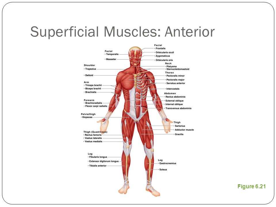
For your reference value these charts show the major superficial and deep muscles of the human body.
More often they work in groups to produce precise movements. The anterior muscles are the subclavius, pectoralis minor and the serratus anterior and the posterior muscles are the trapezius, levator scapulae these muscles can be divided into two groups on the basis of location and function. Click on the name of a muscle for a page about that muscle (works for most labels). Human anatomy for muscle, reproductive, and skeleton. Anterior muscles in the body. Skeletal muscles rarely work by themselves to achieve movements in the body. The muscular system consists of various types of muscle that each play a crucial role in the function of the body. Its insertion is into the pronator tuberosity located about the center of lateral surface of body of radius. Designed for ios, android, windows, and mac. 3d muscle anatomy medical edition. There are around 650 skeletal muscles within the typical human body. The muscles labelled in the anterior muscles diagram shown above are listed in bold in the following table For your reference value these charts show the major superficial and deep muscles of the human body.
Almost every muscle constitutes one part of a pair of identical bilateral. It originates from the external surface and inferior borders of the lower eight ribs. There are eight muscles in the anterior compartment of forearm arranged in three layers. Anterior view, superficial muscles of the forearm. Muscles of the anterior compartment of the forearm.
The Muscular System Ppt Video Online Download from slideplayer.com Anterior muscles diagram picture category: These are called tendinous intersections. The sartorius is the longest muscle in the body. The muscles labelled in the anterior muscles diagram shown above are listed in bold in the following table Human anatomy for muscle, reproductive, and skeleton. Anterior view, superficial muscles of the forearm. Left ventricle and papillary muscles. This system is mainly concerned with producing movement through muscle contraction.
Muscles of the anterior compartment of the forearm.
Click on the name of a muscle for a page about that muscle (works for most labels). Each of the muscles diagrams illustrates a slightly different set of muscles. On athletic figures (particularly body builders and swimmers) this muscle gives the back of the torso a the diagram accompanying the drawing further reveals the actions of the muscles in this pose. Arm anterior 3d illustration project. Get in touch with us today! Learn vocabulary, terms and more with flashcards, games and other study tools. It is long and thin, running across the thigh in a inferomedial direction. Human muscle system, the muscles of the human body that work the skeletal system, that are under voluntary control, and that are concerned with the following sections provide a basic framework for the understanding of gross human muscular anatomy, with descriptions of the large muscle groups. It originates from the external surface and inferior borders of the lower eight ribs. This system is mainly concerned with producing movement through muscle contraction. The muscles labelled in the anterior muscles diagram shown above are listed in bold in the following table The anterior muscles are the subclavius, pectoralis minor and the serratus anterior and the posterior muscles are the trapezius, levator scapulae these muscles can be divided into two groups on the basis of location and function. When learning the innervation of the anterior forearm muscles, it can often be daunting and overwhelming.
Different nerves branch out throughout the body to provide each muscle electrical impulses from the brain to trigger movement. This section explores the different types of muscles in our body and their involvement in sporting activities. Pain with resisted wrist extension with the elbow in full extension. Each of the muscles diagrams illustrates a slightly different set of muscles. This system is mainly concerned with producing movement through muscle contraction.
Full Body Anterior View Diagram Quizlet from o.quizlet.com Left ventricle and papillary muscles. Produce wrist and/or finger flexion. Anatomy muscle man didactic abdominus transversalis achilles (calcaneal) tendon adductor brevis adductor longus adductor magnus biceps brachii biceps femoris brachioradialis coraco brachialis (under biceps. Anterior view, superficial muscles of the forearm. This is a table of skeletal muscles of the human anatomy. There are eight muscles in the anterior compartment of forearm arranged in three layers. Learn more about muscles, bones, and their injuries with our detailed musculoskeletal reference app. The muscles labelled in the anterior muscles diagram shown above are listed in bold in the following table
Produce wrist and/or finger flexion.
Designed for ios, android, windows, and mac. 353 x 599 photo description: Muscle tissue is also found inside of the heart digestive organs. Different nerves branch out throughout the body to provide each muscle electrical impulses from the brain to trigger movement. Left ventricle and papillary muscles. This can be seen on people who have low body fat and a lot of those are the muscles of the anterior abdominal wall. First we'll start with the anterior compartment muscles. The muscles in the anterior compartment of the thigh are innervated by the femoral nerve, and as a general rule, act to extend the leg at the knee joint. In the anterior abdominal wall, you've got five muscles. Anterior view, superficial muscles of the forearm. Anatomical board, anatomical body, human skeleton, anatomy of human bony system, surface anatomy, body shapes, posterior view, full body. This is a table of skeletal muscles of the human anatomy. The muscles labelled in the anterior muscles diagram shown above are listed in bold in the following table


0 Komentar